Introduction to Animal and Plant Cell Coloring Packets

Yo, Medan peeps! Let’s dive into the awesomeness of these animal and plant cell coloring packets. They’re not just for kids doodling – they’re a seriously cool way to learn about the tiny building blocks of life! Think of it as a fun, engaging approach to understanding cell biology, making learning less of a chore and more of a creative adventure.These coloring packets are designed to make learning about animal and plant cells super accessible and enjoyable.
Visual learning is key, and what better way to absorb information than by actively coloring and labeling the different parts of a cell? It’s a hands-on experience that helps solidify understanding in a way that simply reading a textbook can’t. The act of coloring helps memory retention and allows for a deeper understanding of the cell’s structure and function.
Eh, so you’re into that animal and plant cell coloring packet, huh? Pretty geeky, but hey, I get it. Need a break from all those chloroplasts and mitochondria? Check out these anime coloring sheets free for a bit of a chill sesh. Then, you can totally get back to mastering those cell diagrams, man.
It’s all about balance, tau!
Age Appropriateness and Complexity
The beauty of these coloring packets lies in their adaptability. We can tailor the complexity to suit different age groups. For younger learners (say, elementary school), the packets might focus on basic structures like the cell membrane and nucleus, using simple diagrams and labels. Older kids (middle and high school) can tackle more intricate details, including organelles like mitochondria, chloroplasts (in plant cells), and the Golgi apparatus.
We can even incorporate more advanced concepts like cellular processes for the older age group, making it a progressive learning tool. For example, a packet for younger children might feature large, clearly labeled diagrams, while a packet for older children could include more detailed illustrations with subtle differences in organelles highlighted. This ensures that everyone gets a challenging yet manageable learning experience.
Components of Animal and Plant Cells

Yo, Medan peeps! Let’s dive into the nitty-gritty of animal and plant cells. Think of them as the basic building blocks of life, but with some seriously cool differences. We’re gonna break down the key players – the organelles – and see how they work together to keep these tiny powerhouses running smoothly. It’s like comparing two different, equally awesome,
warungs* – both serve a purpose, but have their own unique specialties.
Organelle Comparison: Animal vs. Plant Cells
Alright, let’s get organized. This table shows the major organelles found in both animal and plant cells, highlighting their similarities and differences. Think of it as a cell-based
menu* comparing two different restaurants.
| Organelle | Animal Cell | Plant Cell | Key Differences |
|---|---|---|---|
| Cell Membrane | Present; regulates what enters and exits the cell. | Present; regulates what enters and exits the cell. | Similar structure and function in both. |
| Cytoplasm | Present; jelly-like substance filling the cell, containing organelles. | Present; jelly-like substance filling the cell, containing organelles. | Similar composition and function. |
| Nucleus | Present; contains DNA, controls cell activities. | Present; contains DNA, controls cell activities. | Similar structure and function. |
| Mitochondria | Present; powerhouse of the cell, produces ATP (energy). | Present; powerhouse of the cell, produces ATP (energy). | Similar structure and function, but plant cells may have more due to photosynthesis. |
| Ribosomes | Present; synthesize proteins. | Present; synthesize proteins. | Similar structure and function. |
| Endoplasmic Reticulum (ER) | Present; network of membranes involved in protein and lipid synthesis. | Present; network of membranes involved in protein and lipid synthesis. | Similar structure and function. |
| Golgi Apparatus | Present; modifies, sorts, and packages proteins. | Present; modifies, sorts, and packages proteins. | Similar structure and function. |
| Lysosomes | Present; breaks down waste materials. | Rarely present; plant cells use vacuoles for similar functions. | Plant cells primarily rely on vacuoles for waste breakdown. |
| Vacuoles | Present; small, temporary storage sacs. | Present; large, central vacuole for storage and turgor pressure. | Plant cells have a much larger, central vacuole. |
| Chloroplasts | Absent | Present; conduct photosynthesis, converting light energy into chemical energy. | Unique to plant cells, enabling photosynthesis. |
| Cell Wall | Absent | Present; rigid outer layer providing support and protection. | Provides structural support and protection, absent in animal cells. |
Detailed Organelle Descriptions
Now for the detailed lowdown on each organelle. Think of this as the
special instructions* section of our cell recipe.
The cell membrane is like the bouncer at a club, selectively letting things in and out. The cytoplasm is the cell’s bustling interior, a jelly-like substance where all the action happens. The nucleus is the control center, housing the cell’s DNA, the blueprint for everything. Mitochondria are the powerhouses, generating energy through cellular respiration – it’s like the generator supplying electricity to the whole cell.
Ribosomes are the protein factories, building the proteins needed for the cell’s functions. The endoplasmic reticulum (ER) is a network of membranes involved in protein and lipid synthesis and transport. The Golgi apparatus acts as the post office, modifying, sorting, and packaging proteins for delivery. Lysosomes (in animal cells) are the recycling centers, breaking down waste materials. Plant cells use vacuoles for storage and maintaining turgor pressure – think of it as a water balloon keeping the plant firm.
Finally, chloroplasts (in plant cells) are the solar panels, capturing light energy for photosynthesis, the process that makes plants’ food. The cell wall (in plant cells) is a rigid outer layer providing structural support and protection, like a strong shell.
Illustrative Representation of Organelles
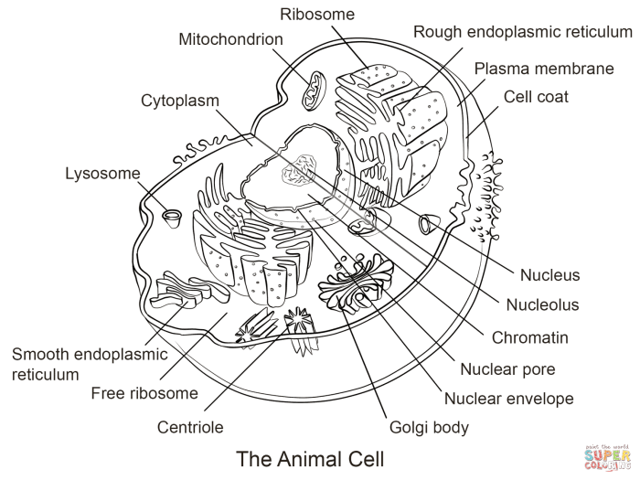
Imagine a typical animal cell as a round, squishy blob. The nucleus sits near the center, like a boss in an office. Mitochondria are scattered throughout, like tiny power generators. The ER forms a network of interconnected tubes and sacs, running throughout the cytoplasm. The Golgi apparatus is near the nucleus, looking like a stack of pancakes.
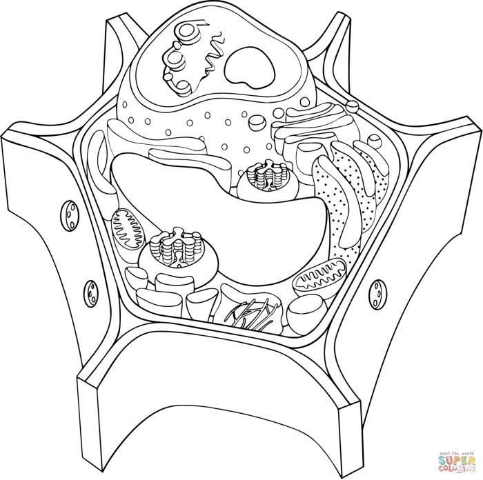
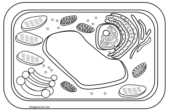
Small vacuoles are scattered around.Now picture a plant cell. It’s more rectangular, thanks to the rigid cell wall. A large, central vacuole dominates the cell, taking up much of the space. The nucleus is still near the center, but the chloroplasts are also visible, scattered throughout the cytoplasm, giving it a greenish hue. The other organelles are present, but the large vacuole and cell wall are the defining features.
Think of it like comparing a simple
- nasi goreng* (animal cell) with a
- nasi uduk* (plant cell) – both delicious, but distinctly different.
Designing the Coloring Packet

Alright, Medan style, let’s get this coloring packet looking
- sick*. We’ve already covered the basics of animal and plant cells, so now it’s time to bring those bad boys to life with some creative coloring fun! Think vibrant colors, clear labeling, and a design that’s both educational and engaging. We want kids (and even some adults!) to actually
- want* to color these cells.
Animal Cell Coloring Page Design, Animal and plant cell coloring packet
For the animal cell, we’re going for a playful, almost cartoonish vibe. Imagine a round, bubbly cell, like a bouncy ball but way more interesting. The nucleus will be a big, bright circle in the center, maybe a sunny yellow or a cheerful orange. We’ll use a slightly darker shade of the same color for the nucleolus, nestled within the nucleus.
The mitochondria, the powerhouses of the cell, will be depicted as smaller, vibrant red bean-shaped structures scattered throughout. The endoplasmic reticulum will be a network of interconnected, light blue squiggly lines, while the Golgi apparatus can be represented by a stack of flattened, slightly curved, purple sacs. Ribosomes, tiny and numerous, will be small, dark green dots scattered around the cytoplasm.
Lysosomes, the cell’s recycling centers, can be depicted as small, bright orange circles. Finally, the cell membrane, the boundary of the cell, will be a thin, dark blue line outlining the entire cell. Labels will be placed neatly beside each organelle, using a simple, easy-to-read font in black.
Plant Cell Coloring Page Design
The plant cell coloring page will be a bit more structured, reflecting the rigid nature of plant cells. We’ll use a rectangular shape to represent the cell wall, with a light brown or beige color. Inside, the cell membrane will be a thin, dark green line. The large central vacuole will dominate the space, a light purple, almost translucent, shape taking up a significant portion of the cell.
The chloroplasts, responsible for photosynthesis, will be depicted as numerous, bright green oval shapes scattered throughout the cytoplasm. We’ll maintain the same color scheme for the nucleus, mitochondria, endoplasmic reticulum, Golgi apparatus, ribosomes, and lysosomes as in the animal cell, to maintain consistency and aid in comparison. The cell wall will be clearly labeled, highlighting its presence as a key difference from the animal cell.
Labels will be similarly placed and styled as in the animal cell design.
Color Key
To ensure clarity and consistency, a color key will be included. This key will list each organelle and its corresponding color. It will be presented in a simple, easy-to-understand format. For example:
| Organelle | Color |
|---|---|
| Nucleus | Yellow |
| Nucleolus | Dark Yellow |
| Mitochondria | Red |
| Endoplasmic Reticulum | Light Blue |
| Golgi Apparatus | Purple |
| Ribosomes | Dark Green |
| Lysosomes | Bright Orange |
| Cell Membrane | Dark Blue |
| Cell Wall (Plant Cell Only) | Light Brown |
| Central Vacuole (Plant Cell Only) | Light Purple |
| Chloroplasts (Plant Cell Only) | Bright Green |
